


Watch video of Dr. Kleeman perfom this surgery which gives a clear picture of the Percutaneous Pedicle Screws procedure. Click Here To Watch Video
A spinal fusion is a bond between two vertebrae building a bridge of living bone across the disc space to unite the vertebrae on either side. It is like a slow setting epoxy which takes about 3 months to set up. If one has a broken table leg that is being repaired with epoxy it often helps to apply a plate and screws to hold it together until the epoxy sets up. In a similar fashion pedicle screws and connecting rods are used as an internal splint to support the spine while the fusion is progressing. They give support to prevent settling of the vertebrae and prevent micromotion both of which can be painful. The use of this internal hardware often frees the patient to resume normal activities right away without having to wear a cumbersome body brace.
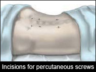

Technology has created a mechanism to insert these screws through small ½" skin incisions. Using sequential dilators, ports are created between the muscles preventing them from being cut and therefore preventing painful scar tissue.


Using radiographic navigation, a diagnostic quality CT scan can be created in the operating room. This scan is then linked to the instruments allowing them to be visualized "real-time" on the CT scan images. The surgery can then be performed through small percutaneous incisions accessing the spine deep inside the body without having to expose it directly.
